


Observing Carrot Organs!
Posted by Joshua Bartosiewicz-
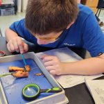
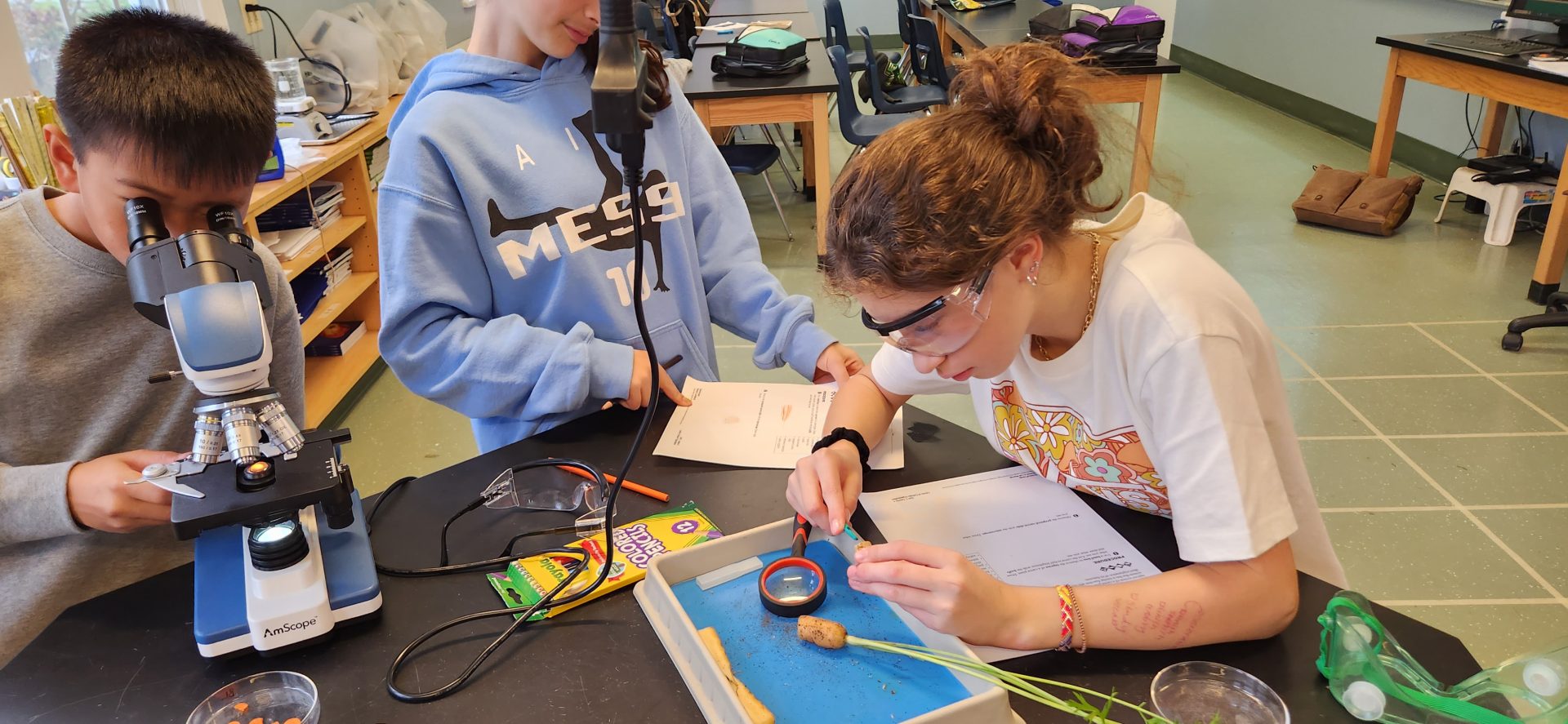
- Slicing up the carrot to observe the organs and parts.
-
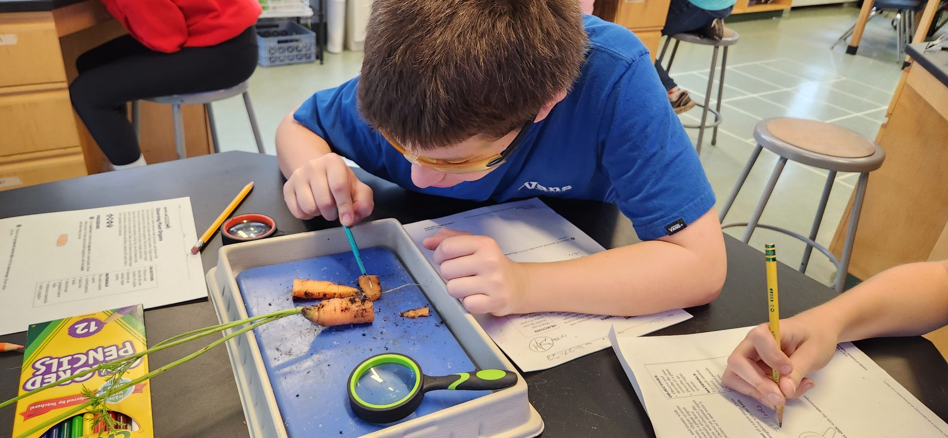
- Cutting up a carrot to see it’s organs.

-
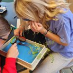
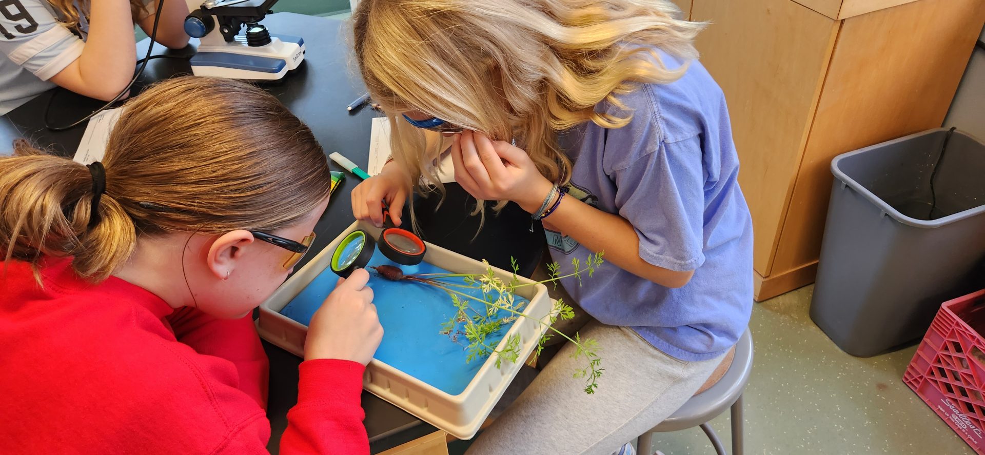
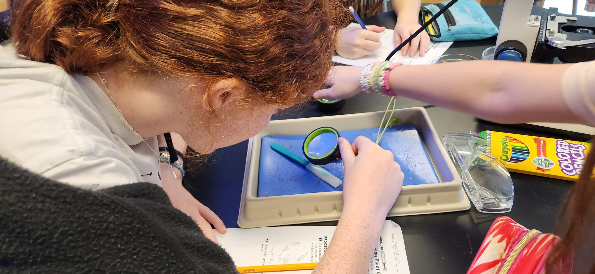
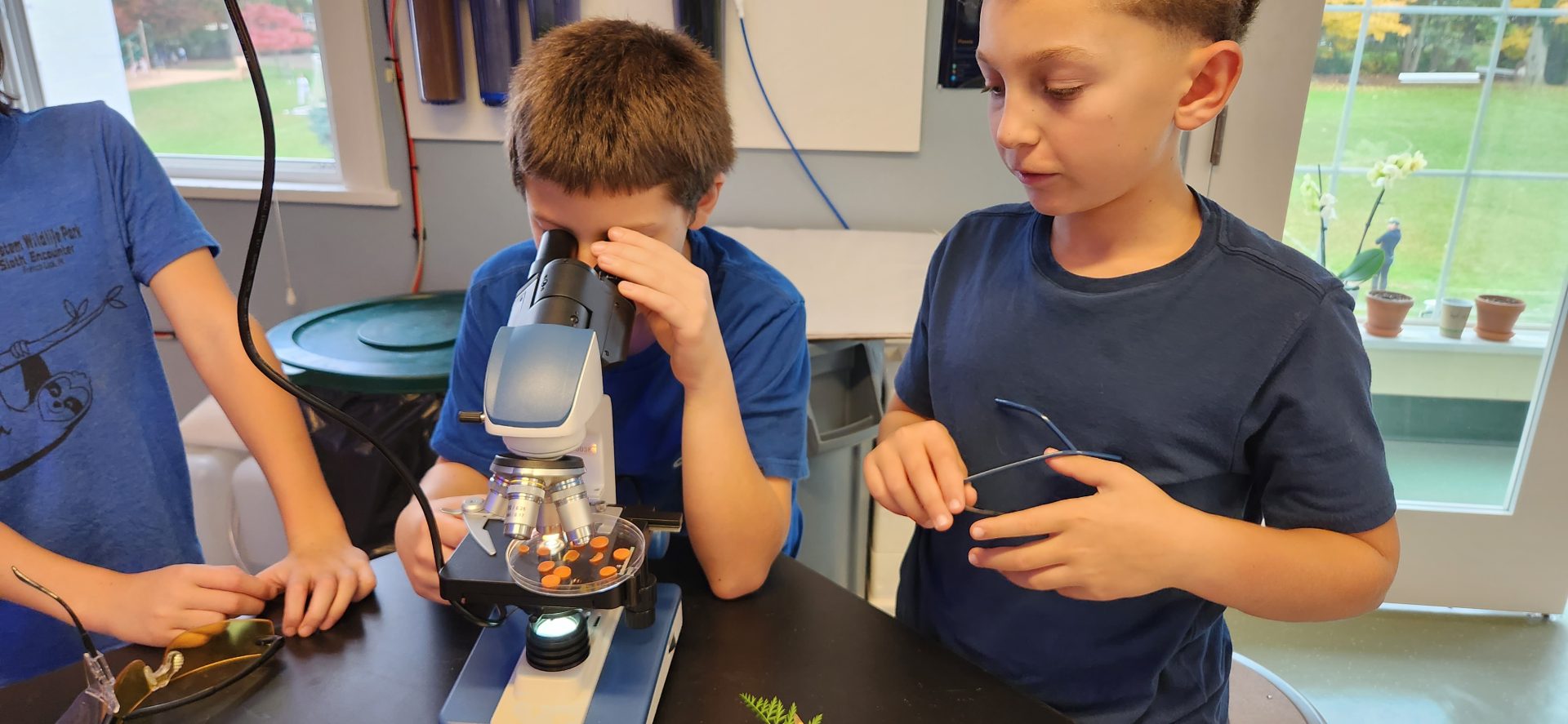
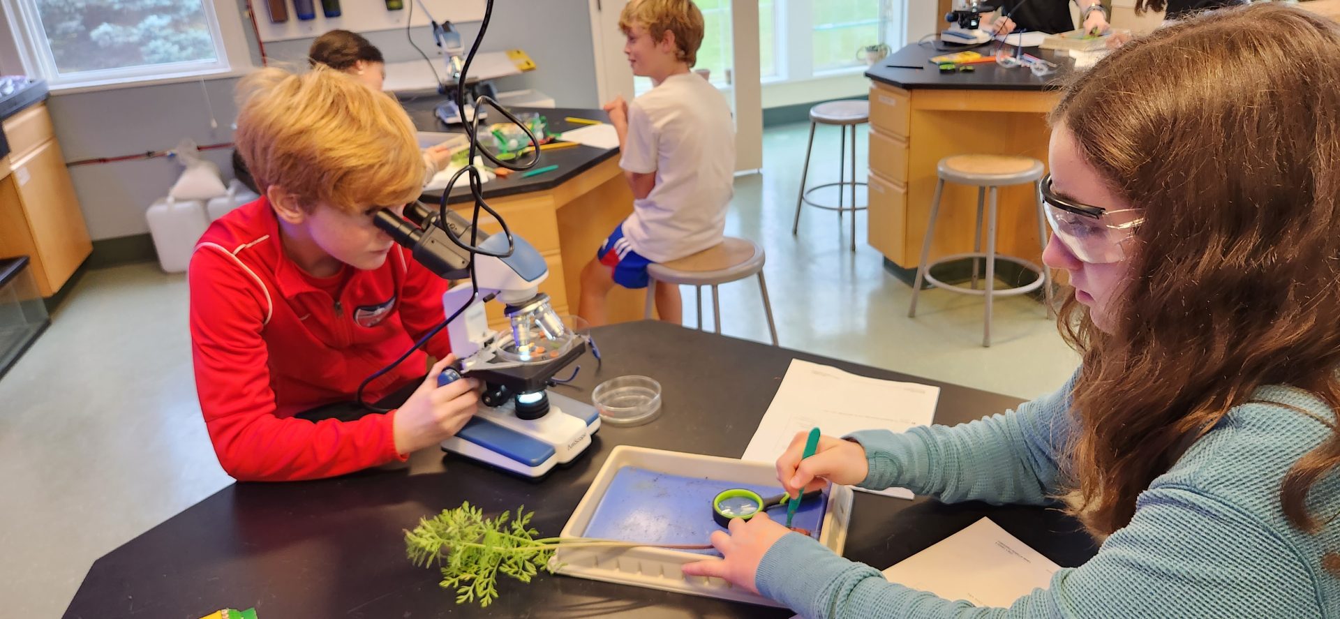
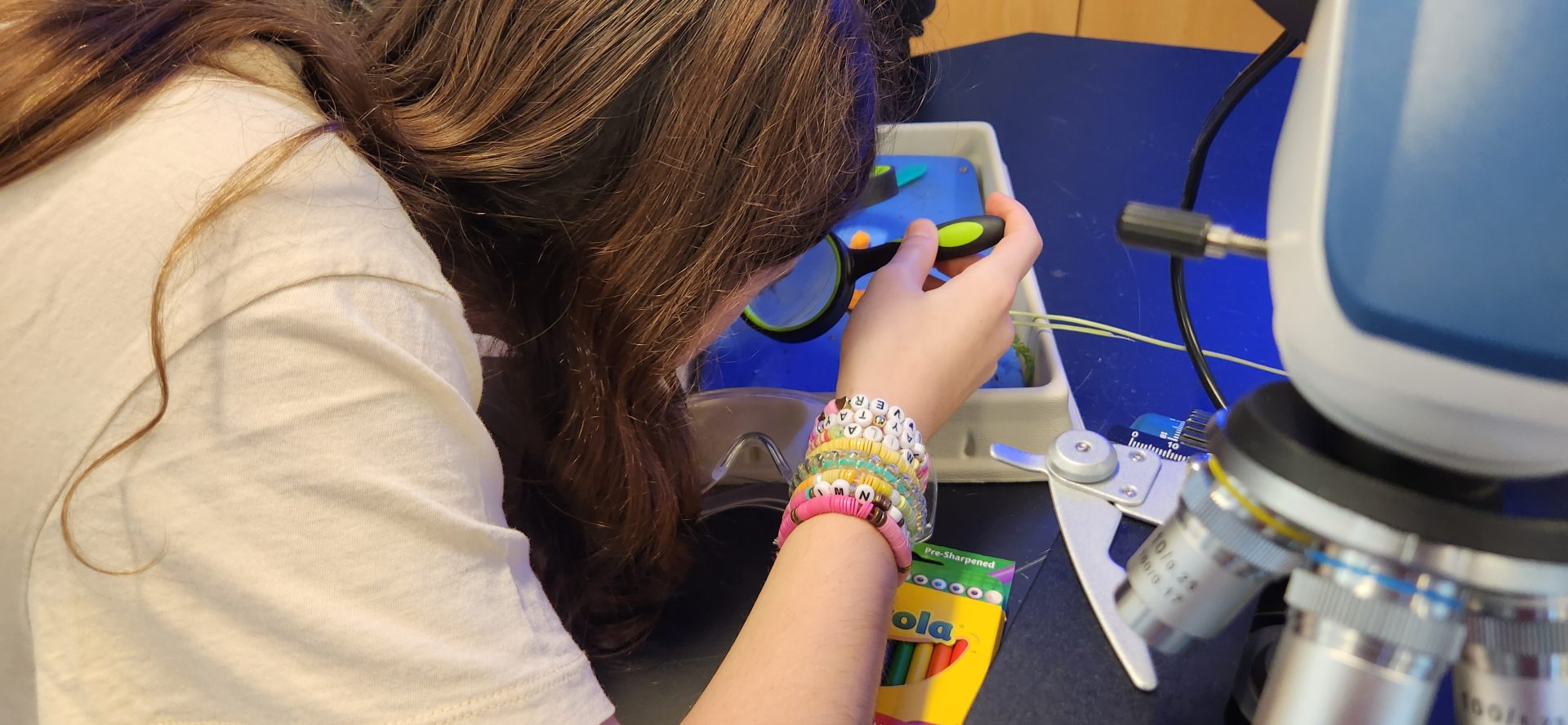
- Checking out the carrot under a magnifying glass.
-
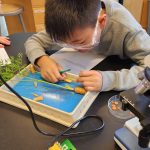
- An even closer look at carrot slices.
-
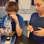
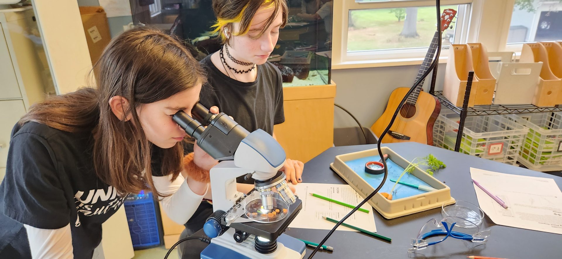
- Observing vascular and ground tissue of a carrot.
-
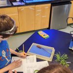
- Recording their observations.

The 7th graders have been studying the levels of organization within different organisms in science. We started out the school year learning about the basic building blocks of life known as cells. Once we learned about cell structures and their functions, we started looking at how these cells work together to help organisms perform life processes. The students began to see that cells are the foundation to organisms, but a collection of similar cells performing a similar task is known as “tissue”. Then, a collection of “tissue” is what makes up our “organs”, from there, a group of organs working together to perform a task are a part of an “organ system”. Finally, a combination of organ systems is what comprises an organism. In conclusion, the levels of organization is as follows; cells -> tissue -> organs -> organ systems -> organisms.
After students were able to grasp that concept, we started looking at the systems that make-up animal bodies (respiratory, circulatory, digestive, muscular systems, etc.) as well as plant bodies (shoot system, root system, etc.). In this lab, the students observed a carrot (which is a taproot to the carrot plant!) with and without a microscope. Our young scholars recorded their observations which included being able to see the vascular tissue and ground tissue of the carrot underneath the microscope. Upon completing this lab, the students were able to see real life examples of the finer details that comprise a plant body (a carrot in this case).
← Our Community's Past Meet Your PE Teacher For the Day, Nina, Grade 2! →